 Local field distribution
around an Au-nanoprism
(Kim et al., OpEx 2012)
Local field distribution
around an Au-nanoprism
(Kim et al., OpEx 2012)
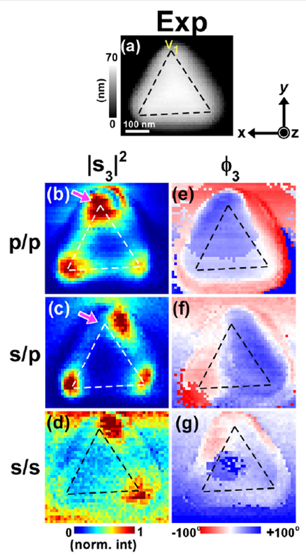
Plasmonics of Nanoparticles: We study the plasmons of individual Ag and Au nanoparticles and their close-coupling with another plasmons. Experimentally, we approach the problem in frequency domain (i. e., spectra), and also in spatial domain (i. e., plasmon modes) to obtain the direct information on plasmon modes and the resonances. For the spatial domain study, in particular, we employ the scattering-SNOM (see Nanoscopy section below) to directly map out the plasmonic local field distribution around such nanoparticles.
- Ahn et al., Phys. Chem. Chem. Phys., 15, 4190 (2013).
- Kim et al., Opt. Express, 20, 8689 (2012).
- Kim et al., Nano Lett. 9, 3619 (2009).
- Kim et al., Opt. Express 16, 1733 (2008).
- Heo et al., ChemComm 46, 6120 (2008).
 Park et al., Nano Lett. 2010
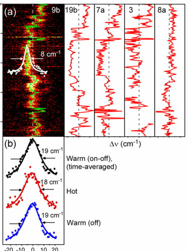
Single-molecule SERS and Beyond: We study the mechanisms behind the surface-enhanced Raman scattering (SERS). By constructing optimized metallic gaps, one can amplify the enhancement and boost up the SERS signals sufficient for the single-molecule detection (sm-SERS). The next step we are currently pursuing is the application of smSERS for the chemical and biochemical reactions that occur at metallic surfaces. The inherent fluctuation in smSERS makes it difficult to differentiate which of the signal components actually carry the genuine chemical information. Our group recently discovered a method to tame the sm-SERS signals, and to differentiate genuine kinetics information from one or a few products. We are extending this proof-of-principle result to the general class of surface reactions of individual molecules at or near the metallic surfaces.
- Choi et al., J. Phys. Chem. Lett. 4, 1079−1086 (2013).
- Park et al., Nano Lett. 10, 4040-4048 (2010).
- Park et al., ChemPhysChem 9, 2491-2494 (2008).
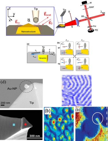
Optical and IR Nanoscopy: In most cases, conventional optical microscopy does not reveal full nano-resolution desired for studying nanostructures and individual molecules. Rather, it only allows us to see images that are significantly blurred. This is called the Abbe's diffraction limit of light. This limit is, however,not a fundamental rule and several techniques exist that evades such limit.
We are developing a nanoscopy technique that enables us to image and spectroscopically characterize nano-objects and individual molecules. In particular, we are developing the scattering-type scanning near-field microscopy (s-SNOM) technique in order to achieve nanometric spatial resolution and full spectroscopic information. The biggest advantage of the s-SNOM is that it can be, in principle, applied to far-IR as well as visible radiation. The aim of this project is to push the limit of the s-SNOM to an individual molecule, particularly in IR and FIR regime, which may lead us to the ultimate goal of IR vibrational spectroscopy of single molecules.
We are developing a nanoscopy technique that enables us to image and spectroscopically characterize nano-objects and individual molecules. In particular, we are developing the scattering-type scanning near-field microscopy (s-SNOM) technique in order to achieve nanometric spatial resolution and full spectroscopic information. The biggest advantage of the s-SNOM is that it can be, in principle, applied to far-IR as well as visible radiation. The aim of this project is to push the limit of the s-SNOM to an individual molecule, particularly in IR and FIR regime, which may lead us to the ultimate goal of IR vibrational spectroscopy of single molecules.
- Kim et al., Opt. Express, 20(8), 8689-8699 (2012).
- Kim et al., Nano Lett. 9(10) 3619 (2009).
- Kim et al., Adv. Mater. 21, 1238, (2009).
- Heo et al., ChemComm 46, 6120 (2008).
- Kim et al., Opt. Express 16, 1733 (2008).
- Kim et al.,Nano Lett.7, 2258 (2007).
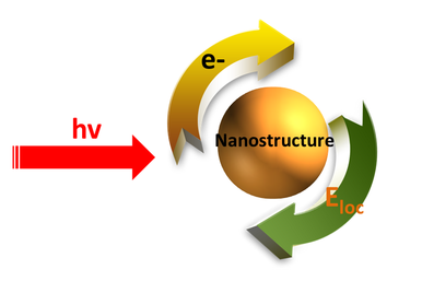
Plasmonic Electrons: the localized plasmons are the coupled oscillation of localized electric field and the oscillating electrons, yet only the localized field has been utilized thus far (mostly field-enhanced spectroscopy). We are currently designing an experiment that probes how the plasmonic electrons behave.
